INDICATIONS AND CONTRAINDICATIONS FOR CHEST CT SCAN
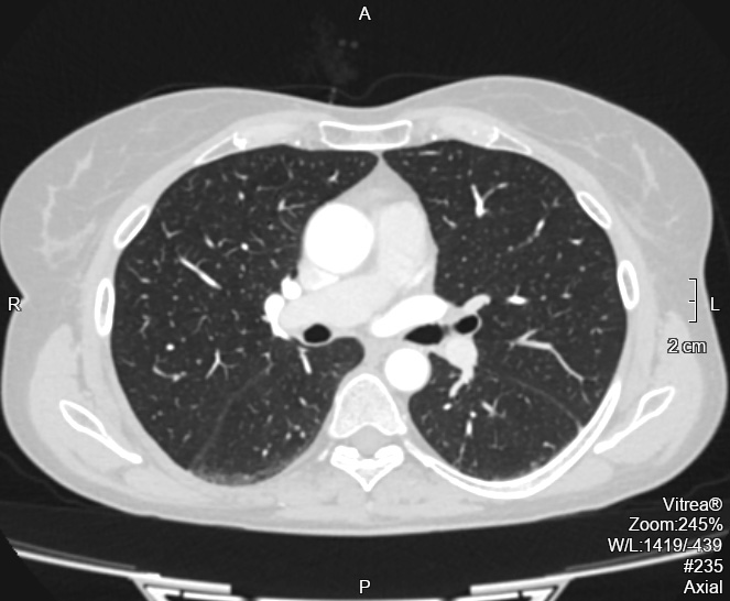
This diagnostic method is used for clarifying and confirming a diagnosis, studying the extent and nature of various injuries, and specifying treatment tactics and details. A chest CT scan in Lviv allows for high-resolution visualization of bone, cartilage, parenchymal, and connective tissue structures of the chest, examining cavities and their contents, blood vessels, the lymphatic system, and the breasts in women. The chest contains many organs and structures, thus there's a wide range of indications for examination using a CT scanner.
A doctor might prescribe a chest CT scan in cases of persistent cough, shortness of breath, chest pain, or suspicion of cancer.
CONTRAINDICATIONS FOR CHEST CT SCAN
- Pregnancy at any stage
- Allergic reactions in patients to iodine-containing contrast agents, hyperthyroidism.
- Diabetes mellitus.
- Renal insufficiency.
Chest CT scans are most commonly prescribed for detecting and treating:
- Infectious diseases and inflammatory processes (pneumonia, lung abscess, pleuritis, tuberculosis);
- Congenital developmental anomalies;
- Benign and malignant tumors in the lungs, mediastinum, and breasts;
- Enlarged lymph nodes at the lung roots and in the mediastinum;
- Pus, blood, or serous fluid in the pleural cavities and the presence of air in the pleural cavity.
HOW A CHEST CT SCAN IS PERFORMED IN LVIV
At the NOVO Medical Center, examinations are conducted by appointment with a referral from a doctor specifying the preliminary diagnosis.
No special preparation is needed for a chest CT scan in Lviv.
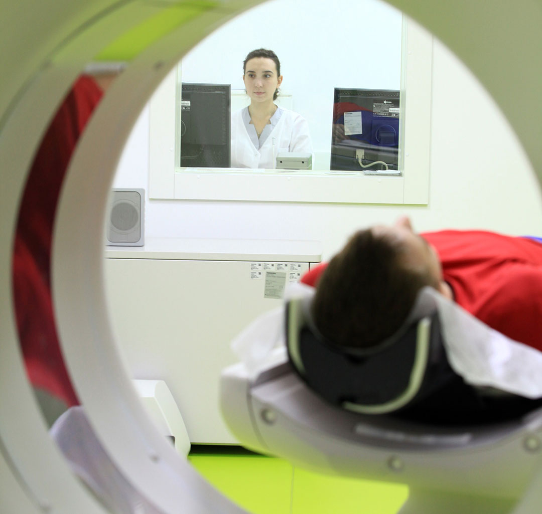
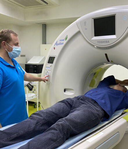
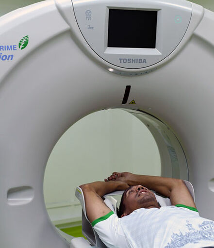
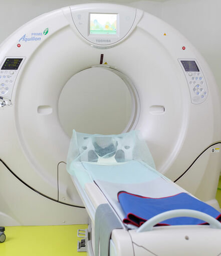
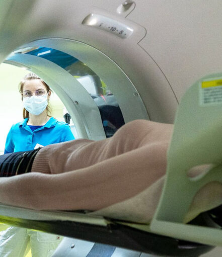
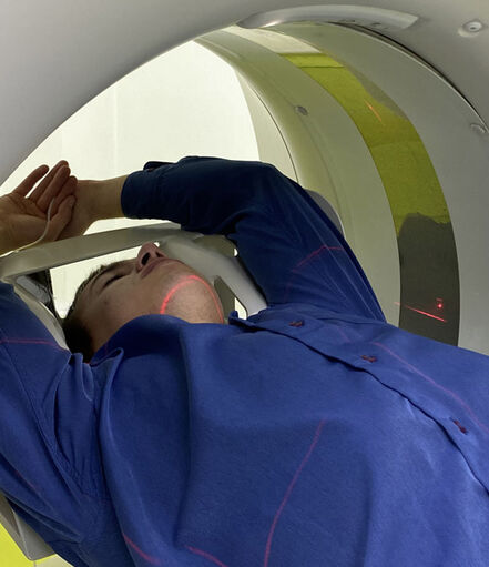
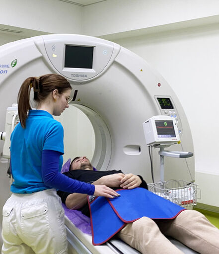
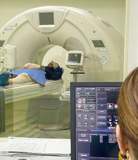
The patient lies on the movable table of the CT scanner, which gradually enters the scanner's opening. Medical staff leave the room where the scanner is located, but the doctor observes the entire scanning process through glass. The patient can communicate with the doctor using intercom communication that works during the scan. The patient might be asked to hold their breath for a few seconds for clearer images.
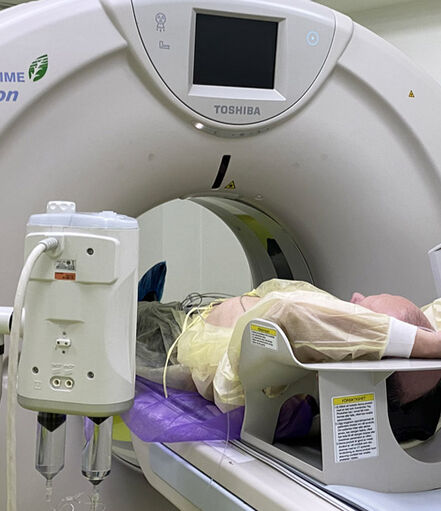
For scans with contrast enhancement, a catheter is inserted into the patient's elbow vein, and through it, using an injector (a special dosing device), the necessary amount of radiocontrast agent is administered.
The entire process takes on average 15-30 minutes.
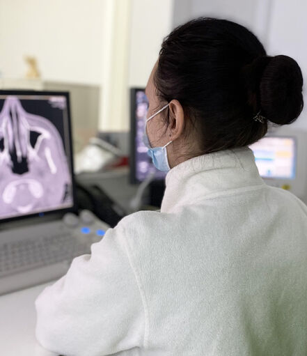
After the scan is completed, the catheter is removed from the elbow vein, the injection site is treated, and the patient returns to their normal routine. The radiology technician reviews all scanned material, prints the most informative data on film, and records the entire scanning process onto a disc. The radiologist then prepares a written report.
